Mission Veterinary Emergency & Specialty

Fluoroscopy
What is Fluoroscopy and How Does It Work?
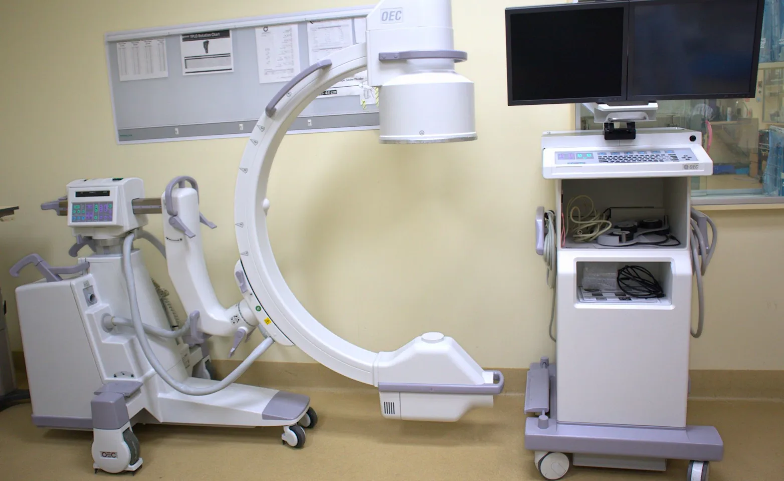
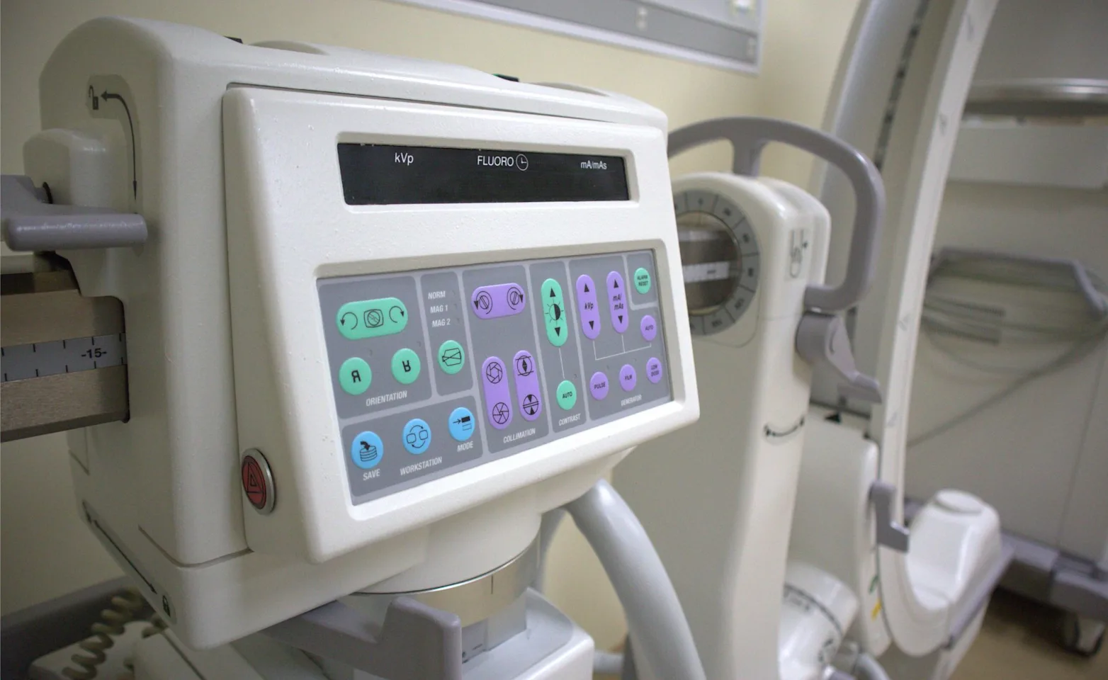
Fluoroscopy is an advanced imaging technique that provides real-time, moving X-ray images of your pet’s internal structures. Unlike standard X-rays, which capture a single static image, fluoroscopy allows our specialists to observe dynamic processes—such as swallowing, breathing, or joint movement—in real-time. This technology is invaluable for diagnosing conditions that require continuous imaging rather than a single snapshot.
During the procedure, a low-dose X-ray beam is passed through the body, displaying the images on a monitor. Depending on the study, your pet may receive a contrast agent to enhance the visibility of specific organs or structures.

Conditions Fluoroscopy Helps Diagnose
Fluoroscopy is a powerful diagnostic tool used to evaluate a variety of conditions, including:
Respiratory Conditions: Tracheal collapse, airway abnormalities, and swallowing disorders
Gastrointestinal Issues: Esophageal strictures, motility disorders, and foreign body obstructions
Orthopedic Concerns: Joint instability, dynamic joint movement assessment, and ligament integrity
Cardiovascular Studies: Vascular abnormalities, dynamic heart function, and congenital defects
Urinary Tract Conditions: Urethral or bladder abnormalities and obstructions
Benefits of Fluoroscopy Compared to Traditional Imaging
Real-Time Diagnosis: Unlike standard X-rays, which provide a single static image, fluoroscopy allows our veterinarians to assess movement and function instantly.
Minimally Invasive: Many fluoroscopic procedures can replace exploratory surgeries or provide critical information for treatment planning.
Enhanced Accuracy: The ability to visualize movement helps diagnose conditions that may not be evident on traditional radiographs or ultrasound.
Guided Interventions: Fluoroscopy is often used to assist with minimally invasive procedures, such as stent placements or joint injections.

Specialists and Departments Utilizing Fluoroscopy
Fluoroscopy is a valuable tool across multiple veterinary specialties, including:
Emergency & Critical Care: Diagnosing airway obstructions, foreign body ingestion, or dynamic heart and lung conditions
Internal Medicine: Evaluating gastrointestinal motility, tracheal function, and vascular abnormalities
Surgery: Assisting in orthopedic repairs, joint assessments, and minimally invasive procedures
Cardiology: Examining real-time cardiac function and vascular flow
Neurology: Assessing spinal abnormalities and guiding interventional procedures
Q: Is fluoroscopy safe for my pet?
A: Yes! Fluoroscopy uses low-dose radiation, and our team takes all necessary precautions to minimize exposure. The benefits of real-time imaging far outweigh any minimal risks associated with radiation exposure.
Q: Will my pet need sedation for a fluoroscopy?
A: It depends on the type of study. Some procedures require light sedation to keep your pet comfortable and still, while others can be performed with minimal restraint.
Q: How long does a fluoroscopy procedure take?
A: Most fluoroscopic studies take between 20–45 minutes, depending on the complexity of the assessment.
Q: Can my pet eat before the procedure?
A: Fasting requirements depend on the study. For gastrointestinal imaging, your pet may need to fast for 8–12 hours beforehand. We will provide specific instructions based on your pet’s needs.
Q: How do I schedule a fluoroscopy appointment?
A: Referring veterinarians can send a referral through our online portal, or pet owners can contact our hospital directly. Our team will work with you to determine if fluoroscopy is the right diagnostic tool for your pet.
